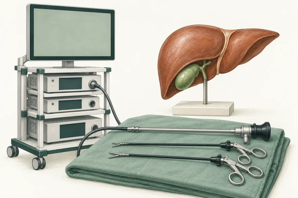
Intraoperative cholangiography, often shortened to IOC, is an X-ray examination of the bile ducts performed during an operation. During gallbladder removal, it can help the surgeon understand the duct anatomy, look for stones and investigate suspected injury. It is an additional source of information, not a guarantee that every problem will be detected or prevented.
How is the examination performed?
While you are under general anaesthesia, the surgeon usually places a thin catheter into the cystic duct, the small duct connected to the gallbladder. Contrast liquid is introduced and moving X-ray images, called fluoroscopy, show its passage through the biliary system. The team assesses the duct outlines and whether contrast reaches the small intestine.
This is imaging performed within the operation, rather than an endoscope passed through the mouth. The basic steps are described in Cleveland Clinic’s patient information. A cholangiogram does not itself remove a stone: treatment requires an additional manoeuvre or procedure.
What questions can IOC help answer?
- Are the ducts connected as expected, or is there an anatomical variation?
- Is there a finding suggesting a stone or obstruction in the common bile duct?
- If injury is suspected, does the image help establish what has happened?
The reason may be apparent before surgery, for example because tests suggest a duct stone, or arise during the operation when anatomy is difficult to interpret. The multi-society safe cholecystectomy guideline particularly supports intraoperative biliary imaging when anatomy is uncertain or bile duct injury is suspected. Read more about the underlying condition in our guide to common bile duct stones.
Is cholangiography necessary for everyone?
Practice varies. The 2025 SAGES imaging guideline conditionally favours IOC, including routine rather than selective use in adults undergoing laparoscopic gallbladder removal for benign disease. The certainty of the supporting evidence is low. “Conditional” is important: it does not mean every patient must have this test regardless of circumstances.
A subsequent SAGES clarification emphasises individual decisions, surgical experience, patient factors and available resources. With normal tests, clearly identified anatomy and no additional indication, different choices can be reasonable. Ask how your team selects a method and which options are available; no article can confirm equipment or routine practice at a particular hospital.
How does IOC differ from other tests?
MRCP
Magnetic resonance cholangiopancreatography is a specialised MRI examination. It produces images without X-rays and without inserting a catheter into the bile duct. It is commonly used outside the operation to investigate suspected duct problems. It shows anatomy but does not remove stones.
ERCP
Endoscopic retrograde cholangiopancreatography uses an endoscope passed through the mouth to reach the opening of the bile duct in the intestine. It combines endoscopy and X-ray imaging and can remove stones or place a stent. Because it has its own risks, it is generally used when treatment is expected, not simply as a replacement for every imaging test.
Fluorescence imaging and ultrasound
Indocyanine green, or ICG, fluorescence uses a dye and a special near-infrared camera to help show biliary anatomy without X-rays. It is not the same as a contrast X-ray cholangiogram and cannot be assumed to answer the same questions about duct stones. Laparoscopic ultrasound is another option in trained hands. Their suitability depends on the clinical question, equipment and expertise.
What if the image is abnormal or unclear?
A suspected filling defect may represent a stone, but air bubbles or technical limitations can create misleading appearances. The surgeon assesses image quality and the overall situation before deciding what to do. Further imaging may be needed rather than automatically proceeding to ERCP.
If a stone is confirmed, possible approaches include removal during surgery or ERCP at an appropriate time. Suspected duct injury requires a separate assessment and management plan, sometimes with specialist input. A normal image is reassuring only within its limits and does not cancel the need to investigate concerning symptoms later.
What are the limitations and risks?
IOC adds operating time and involves radiation and contrast. Catheter placement may be difficult, especially with a small or inflamed duct, and a useful image is not always obtained. Potential complications include a contrast reaction, duct injury, bleeding or infection. Interpretation also matters: false-positive findings can lead to unnecessary procedures. The expected benefit should therefore be considered alongside the individual risks.
What should you discuss before and after surgery?
Tell the team about any previous reaction to an injected contrast agent, all medicines and supplements, and pregnancy or the possibility of pregnancy. Follow your own preoperative instructions; do not stop medicines without advice. Ask why imaging may be useful for you and what the plan would be if a stone were found.
After surgery, ask whether IOC was performed, whether the study was complete and whether any follow-up is required. Recovery is mainly determined by the operation and any additional treatment. Worsening abdominal pain, fever, jaundice or persistent vomiting needs prompt medical assessment, even after a normal cholangiogram.
Frequently asked questions
Will I feel the contrast injection?
IOC during gallbladder removal is performed while you are under general anaesthesia. It is not usually a separate awake examination.
Does an abnormal cholangiogram always mean a stone?
No. Image quality and possible artefacts must be considered alongside other findings. The team will explain what is suspected and how it will be checked.
Does IOC replace careful surgical identification of the ducts?
No. It supports anatomical assessment but does not replace safe dissection, surgical judgement or a change of strategy when needed.
Will I need a cholangiogram if MRCP was normal?
Possibly. MRCP and IOC answer partly overlapping questions at different times. New findings or uncertainty during surgery can justify additional imaging.
For a planned discussion of your investigations, contact the surgical team. Bring previous scan reports and describe any contrast reactions. This information does not replace an individual treatment plan.