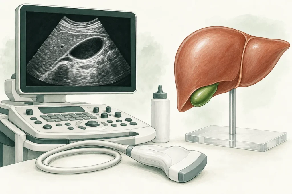
A report describing a “dilated common bile duct” can be worrying, particularly if an ultrasound did not show a stone. The common bile duct carries bile towards the intestine. A larger measurement may reflect an obstruction, but it can also occur without a blockage. The useful question is whether the finding fits your symptoms, blood tests and earlier imaging.
Why one measurement is not enough
There is no single diameter that separates normal from abnormal for every adult. Age, previous gallbladder removal, the imaging method and the place where the duct was measured all affect interpretation. An ultrasound measurement should not be compared mechanically with a number on CT. A stable finding in a person who feels well means something different from new enlargement with jaundice.
After cholecystectomy, non-obstructive enlargement is possible, as described in a study of long-term CT changes. This does not mean that every postoperative enlargement is harmless. New symptoms or other abnormal findings still need assessment. An old scan is useful evidence, not a reason to ignore a change.
What the doctor looks for
The assessment starts with when the scan was done and why. Tell the clinician about pain in the upper abdomen, itching, fever, a change in urine or stool colour, reduced appetite and unintended weight loss. Mention previous gallbladder or bile duct procedures and all medicines, including opioid painkillers. Do not stop a prescribed medicine simply because it might affect the duct.
- The report should identify which ducts are enlarged and whether a stone, narrowing or other lesion is visible.
- Blood tests may include bilirubin, alkaline phosphatase, GGT and liver enzymes. Their pattern and trend matter more than one isolated result.
- Previous imaging helps establish whether the finding is longstanding, newly developed or progressing.
- Symptoms and examination determine whether outpatient investigation is appropriate or urgent hospital assessment is needed.
Possible causes include duct stones, a scar-related narrowing and, less commonly, a growth affecting drainage. Ultrasound does not always show the cause, especially in the lower part of the duct. Neither a negative stone finding nor a normal blood test on one occasion settles every case.
When further tests are considered
In the investigation of suspected gallstone disease, NICE recommends considering MRCP when ultrasound has not shown a duct stone but the duct is enlarged or liver blood tests are abnormal. MRCP uses MRI to examine the bile ducts. Endoscopic ultrasound may help when the diagnosis remains uncertain. These are selected tests, not a checklist everyone must complete.
ERCP is generally reserved for a situation in which treatment, such as removing a stone or restoring drainage, is expected. It is not the automatic next step for an incidental measurement because it carries risks, including pancreatitis. Where the overall assessment suggests a benign, stable finding, a clinician may recommend no additional investigation or an individualized follow-up plan. Ask what would change that plan.
Leave the consultation knowing who will review the next result, whether blood tests need repeating and which changes require an earlier review. If two reports disagree, bring both reports and their images. Differences in the method or measurement location can sometimes explain the discrepancy, but a clinician needs to check it in context.
Symptoms that should not wait
Seek prompt medical assessment for new jaundice, dark urine with pale stools, or persistent upper abdominal pain. Pain accompanied by fever or shaking chills, especially with jaundice, requires urgent hospital assessment because an infected, obstructed bile duct is possible. Confusion, fainting or marked weakness are reasons to seek emergency help. Do not wait for a routine appointment or an outpatient scan when you are acutely unwell.
Preparing for the consultation
Bring the complete ultrasound report and available images, earlier scans, recent blood results with dates, and records of gallbladder surgery or ERCP. Note whether a stent was placed and whether its removal is documented. A short symptom timeline is more useful than trying to interpret the millimetres yourself. Ask: “Is there evidence of impaired drainage, what uncertainty remains, and who will review the next result?”
Frequently asked questions
Does enlargement mean I have cancer?
No. Several benign explanations are possible. Persistent unexplained enlargement or associated concerning findings should be investigated rather than guessed at from the report.
Can a stone be present even if ultrasound did not find it?
Yes. Small stones and parts of the duct may be difficult to see. Further testing depends on the whole assessment.
Do I need an operation if the gallbladder has already been removed?
Not because of the measurement alone. Treatment depends on the cause; some findings need no procedure.
Can diet or a “bile cleanse” reduce the diameter?
A cleanse does not diagnose or safely treat an obstruction. Avoid self-treatment and agree a clear investigation plan.
Read more about MRCP and endoscopic ultrasound. For a planned review of your results, use the contact page. This information supports, but does not replace, an individual medical assessment.