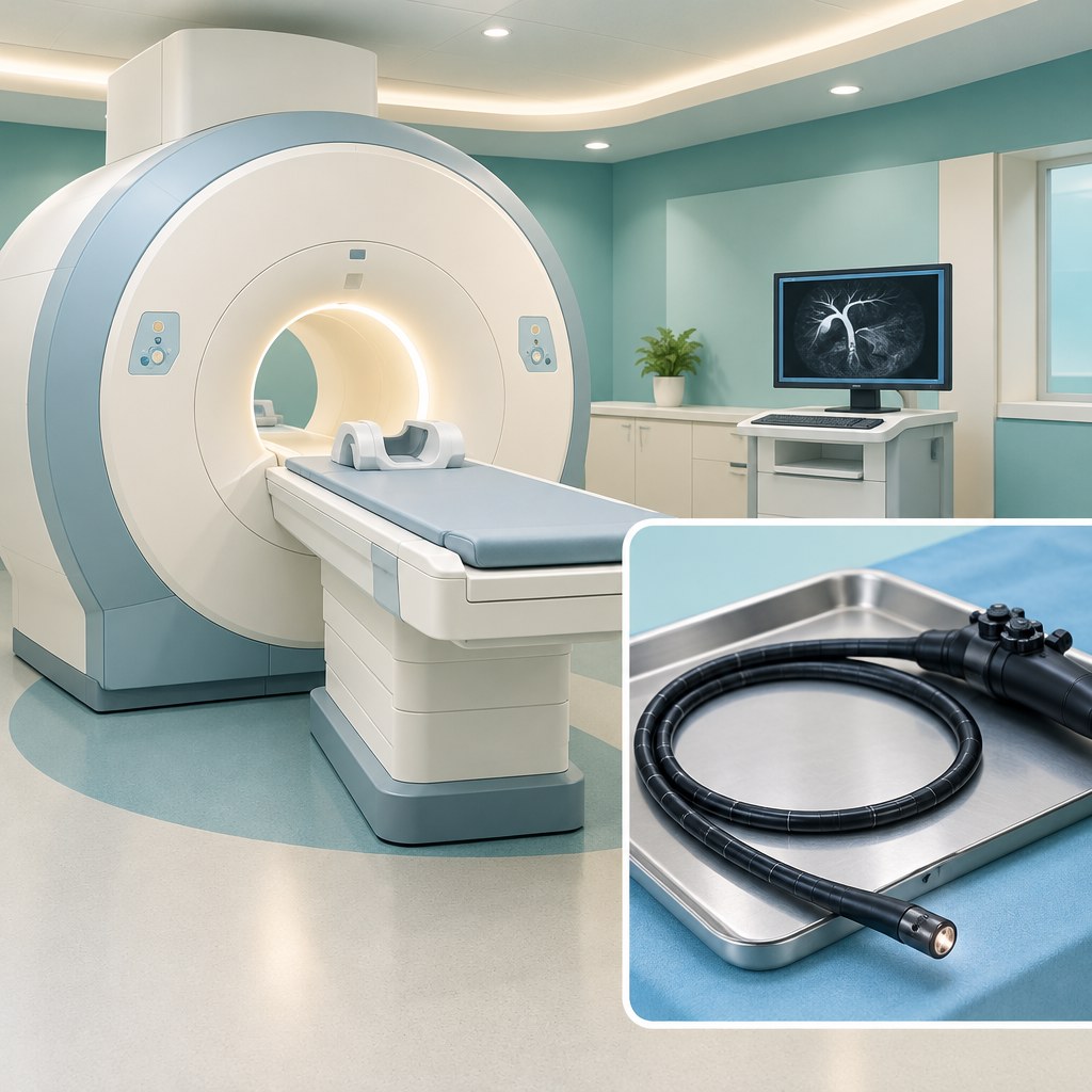
When an ultrasound does not clearly show whether there is a stone in the common bile duct, a doctor may suggest MRCP or endoscopic ultrasound. Both tests help clarify the diagnosis. The choice depends on blood test results, symptoms, possible limitations and the team’s experience. The most expensive or complex test is not necessarily the best one for you.
Why examine the bile ducts specifically?
Stones in the gallbladder and stones in a bile duct are different clinical situations. A stone that has moved into a duct can obstruct the flow of bile. However, a standard ultrasound does not always show the stone directly. A dilated duct or changes in liver blood tests may provide clues.
The doctor assesses these findings together. For an intermediate likelihood of a duct stone, the ASGE guideline allows either MRCP or endoscopic ultrasound. The aim is to obtain enough information for the next decision while avoiding an unnecessary invasive procedure. ASGE recommendations.
MRCP: images of the ducts without an endoscope
Magnetic resonance cholangiopancreatography, or MRCP, is a specialised MRI examination. It shows the bile and pancreatic ducts without X-ray radiation. No endoscope is passed through the mouth. To obtain good-quality images, you need to remain still and follow breathing instructions.
Before booking, tell the team about a pacemaker, other implants, metal fragments, pregnancy or severe claustrophobia. An implant does not always rule out MRI: its precise model and conditions for safe scanning matter. If contrast is planned, ask why it is needed and report kidney disease and any previous reactions to contrast agents. ACR/RSNA information about MRCP.
Endoscopic ultrasound: a closer view
Endoscopic ultrasound, or EUS, uses a flexible endoscope with an ultrasound probe. It is passed through the mouth into the upper digestive tract to examine nearby structures. The method can be useful for finding small stones, particularly when earlier tests have not resolved the question.
Sedation is often used. Follow the fasting instructions and report allergies, other medical conditions and all medicines. Do not stop anticoagulants or diabetes medicines yourself. If sedation is planned, arrange for someone to accompany you home and follow the driving restrictions afterwards.
EUS carries procedural risks, including reactions to sedation and rare injury to the wall of the digestive tract. The risks change if a needle puncture or another intervention is also performed. A biopsy is not usually the aim when simply looking for a stone. NHS information about EUS.
How to choose between the two tests
- First question: what exactly needs to be confirmed or ruled out after ultrasound and blood tests?
- Second: are there limitations to MRI or an increased risk from sedation?
- Third: which examination is available within the required timeframe, and who will interpret it?
- Fourth: how will a positive or negative result change the treatment plan?
Sometimes EUS is needed after MRCP if the diagnosis remains unclear. This does not necessarily mean that the first test was performed incorrectly. The methods have different strengths and limitations. Their accuracy and risks are considered by NICE.
Frequently asked questions
Will a stone be removed during MRCP or diagnostic EUS?
These tests are primarily diagnostic. If a stone is confirmed, the doctor will discuss treatment separately. ERCP is a different procedure that may be used to remove stones from a duct.
Does everyone need both tests?
No. One is often enough. Before a second test is arranged, ask what specific question remains unanswered. Bring previous images, not just a short written report.
If no stones are found, can I forget about the pain?
A negative result does not automatically explain every symptom. Recurrent pain needs reassessment: there may be another cause, and the condition may have changed since the examination.
How should I check the preparation instructions before booking?
Ask for written instructions from the centre performing your test: when to stop eating and drinking, which documents to bring and how to agree on your morning medication. Also clarify whether sedation is planned, who will explain the result and whom to contact after the procedure. Do not reuse instructions from a previous gastroscopy or MRI for a new examination without checking.
When emergency help is needed
Severe or persistent upper abdominal pain, particularly with fever, chills, jaundice or repeated vomiting, requires urgent assessment. Do not wait for a routine MRI if your condition is worsening. Dark urine and very pale stools should also be discussed with a doctor without delay. Warning symptoms according to NIDDK.
What to bring to a consultation
Prepare your ultrasound results, blood test results, previous MRI examinations and medication list. Note when the pain started, how long it lasts and whether you have had a fever. This helps make the discussion of further tests more specific.
For a consultation, call +380953566412. Read more about gallbladder removal and consultation with a surgeon.
This article is for information and does not replace an individual medical examination.
Read also: ERCP to treat bile duct stones and preparing for ultrasound.