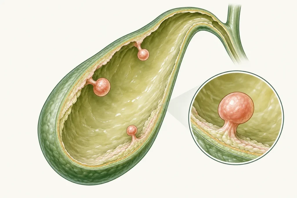
A gallbladder polyp is a projection or lesion on the inner wall of the gallbladder. It may be a true tissue growth or a pseudopolyp, such as a cholesterol deposit. Specific symptoms are usually absent, so lesions are often found incidentally on ultrasound. Not every polyp needs treatment or surgery: management depends on size, appearance, growth and risk factors. When surgery is indicated, the gallbladder is usually removed together with the lesion.
Definition
A polyp projects from the lining into the inside of the gallbladder. It may be rounded, attached by a thin stalk or have a broad base. Most small findings are benign, meaning non-cancerous, although ultrasound cannot always show the exact type of tissue involved. Polyps are found in people of different ages and sexes.
The ICD-10 code is selected according to the established diagnosis and the classification version in use, by the clinician or medical coding team.
Polyps may occur singly or in groups; the term “polyposis” describes multiple polyps. When choosing a treatment plan, the clinician considers their number together with their size, shape and other features. The number alone does not determine whether surgery is needed.
Some true neoplastic polyps have malignant potential, so risk assessment matters. Most small cholesterol pseudopolyps do not carry that risk. Impaired bile drainage is possible in certain circumstances, but most small polyps do not block ducts or disrupt digestion.
Factors associated with polyp formation
The reasons polyps form depend on their type. Cholesterol pseudopolyps are associated with lipid deposits in the gallbladder wall, while inflammatory pseudopolyps develop alongside chronic inflammation. True tumours have a different origin that cannot always be established. Any contribution from inherited factors or metabolic disorders therefore needs to be considered in the context of the particular finding.
Tell the clinician about any other health conditions during your consultation. They help build a picture of your overall health, although not all have a proven link with the development of polyps:
- Endocrine conditions;
- Hormonal disorders;
- Disturbances of lipid metabolism;
- Biliary conditions, including stones, cholangitis or cholecystitis;
- Conditions affecting the immune system.
Balanced meals and avoiding overeating support general health, but eating habits cannot establish the cause of an individual polyp. When the medical history suggests it, the clinician may also consider parasitic infections that can affect the bile ducts. These need their own assessment and do not explain most polyp-like findings.
Types
Lesions can be classified by their nature:
- Cholesterol pseudopolyps. These are associated with cholesterol deposits in the wall, project into the lumen and usually remain fixed when body position changes. Suspecting this type alone does not require immediate surgery;
- Inflammatory pseudopolyps develop alongside inflammation, sometimes associated with stones or another source of irritation to the gallbladder wall. Investigations help identify an accompanying condition, which then guides the choice of treatment;
- Adenomatous polyps are true tissue growths that may have the potential to become cancerous. If this type is suspected, a surgeon assesses the findings and explains whether removal is appropriate;
- Papillary lesions or papillomas are finger-like mucosal growths. Their nature and the need for surgery require individual assessment; histology determines the exact type.
When several polyps are present, the clinician assesses each finding, paying particular attention to the largest. Symptoms, size, shape, growth and individual risk factors guide the decision. Any accompanying stones are also considered when choosing monitoring or surgery, with an explanation of how they affect the recommendation.
Symptoms
Most polyps do not cause characteristic symptoms. If you have pain or digestive problems, the clinician investigates their cause: these complaints can be related to other digestive conditions and, sometimes, to heart or lung disease.
Possible complaints requiring assessment include:
- Nausea and, less often, vomiting;
- Abdominal heaviness;
- Heartburn;
- A bitter taste;
- Dull, sharp or aching pain below the right ribs or in the upper central abdomen.
When bile drainage is obstructed, bilirubin can build up in the blood and cause yellowing of the skin and whites of the eyes. This jaundice needs urgent investigation, even if a previous ultrasound showed a small polyp.
Polyps very often cause no symptoms and are discovered incidentally during ultrasound.
Diagnosis
Assessment usually begins with ultrasound. It can show single or multiple growths attached to the gallbladder wall. A typical polyp stays in place and does not cast an acoustic shadow on the image. Sludge and other changes can sometimes look similar, so the clinician considers the images alongside the rest of the assessment.
Endoscopic ultrasound may help in difficult cases. A flexible endoscope with an ultrasound probe is passed through the mouth to examine the area from close by. It may clarify structure but is not required for every small polyp.
MRI, including magnetic resonance cholangiography, may be used to assess the bile ducts, suspicious lesions or associated changes. It is ordered when indicated and cannot always determine a polyp’s exact histological type.
Polypoid findings must be distinguished from malignant tumours, sludge, stones and changes associated with calculous or acalculous cholecystitis. Parasitic disease may be considered when the history suggests it.
Possible complications
If a lesion has concerning features, keep to the follow-up schedule agreed with your clinician. Some true tissue growths may be precancerous, whereas most small pseudopolyps are benign. A large growth in a particular location can sometimes interfere with bile drainage. The clinician checks its size and position, looks for signs of impaired drainage and explains which changes in symptoms you should watch for.
The assessment also looks for accompanying stones, blocked ducts or severe inflammation. The following conditions may be associated with those problems, rather than being typical direct effects of a small polyp:
- Pancreatitis, including recurrent episodes or chronic pancreatic disease;
- Cholecystitis;
- Gallbladder wall perforation;
- Bile leakage into the abdomen and biliary peritonitis.
A broad-based, or sessile, lesion needs careful assessment, particularly if the nearby gallbladder wall is thickened. The clinician considers this feature together with the polyp’s size and growth, your age and other factors that may affect risk.
Reviewing the findings helps the clinician recommend monitoring or surgery. The discussion should explain the expected benefits of each approach, possible complications and any changes that would prompt a review of the plan.
Treatment
Medicines do not reliably eliminate a true neoplastic polyp. However, many small findings do not need surgery. European guidance uses ultrasound at six months, one year and two years for some lesions; a lesion no larger than 5 mm without risk factors may need no follow-up. A clinician should specify the plan because recommendations differ somewhat between guidelines.
When considering removal of the gallbladder and its polyp, clinicians assess:
- Size: European guidance recommends considering cholecystectomy for lesions measuring 10 mm or more when the patient is fit for surgery and agrees to it;
- Growth: if a lesion increases by at least 2 mm during two years of follow-up, the clinician reassesses its size and other risk factors. The combined findings guide the decision about surgery;
- Malignancy risk, including older age, primary sclerosing cholangitis, suspicious morphology or wall thickening. With certain risk factors, surgery may be considered for lesions measuring 6-9 mm;
- Gallbladder inflammation and associated symptoms when there is no better explanation;
- The number of lesions, considered together with their size and other characteristics, as multiple polyps alone do not determine the need for surgery;
- Confirmed impaired bile drainage and its cause.
When a suspicious lesion requires surgery, the standard approach removes the gallbladder rather than simply cutting off the polyp. The choice depends on the likely lesion type and tumour risk.
The operation is called cholecystectomy. It can be performed laparoscopically or through an open incision.
Before surgery, the team examines you and makes an individual investigation plan. Depending on the clinical question, tests may include ultrasound, CT, X-rays, an ECG and laboratory tests. The clinician should explain the purpose of each test. The full list, including microscopic examination of bile, is not needed for every patient.
Open cholecystectomy uses a larger incision in the abdominal wall, usually leaving a larger scar and requiring a longer recovery. It may be chosen for difficult anatomy, significant inflammation or suspected cancer. For a lesion measuring 15-18 mm, the surgeon also considers its shape, growth and other features. Tissue examination establishes its nature, while the complete assessment guides the surgical approach. If cancer is suspected, a more extensive operation may be needed.
In general, an open approach involves:
- Incising the tissue layers;
- Retracting tissues to access the gallbladder;
- Safely identifying and closing the cystic duct and artery, separating the gallbladder from the liver and removing it;
- Closing the wound in layers.
The operation is performed under general anaesthesia. It may take around two hours, although the complexity of the procedure affects its duration. Your surgeon can explain the expected timing for your situation.
Laparoscopic cholecystectomy uses smaller incisions. Instead of one long incision, which may measure 15-20 cm in open surgery, there are often four openings of approximately 0.5-2 cm. Their number and size can vary. A camera, trocars and surgical instruments are introduced through them.
Carbon dioxide is introduced into the abdomen to create working space. The camera sends images to a monitor so the surgeon can guide the procedure.
The operation is performed under general anaesthesia and often takes around an hour, with more time needed in some complex cases. Recovery is usually easier than after an open operation. Before surgery, the surgeon discusses possible complications, including bleeding, infection, a postoperative hernia and injury to the bile ducts.
Most people can lead a full life after gallbladder removal. Bile reaches the intestine without intermediate storage, so tolerance of food may change during the first weeks. A lifelong special diet is not necessary for everyone; healthy eating and individual advice are usually appropriate.
If the findings suggest a cholesterol pseudopolyp, the clinician assesses its risk and whether monitoring is needed. Long-term bile acid medication is not a reliable way to dissolve the lesion. Medicines for pain, infection, inflammation or impaired bile drainage may be useful for associated problems, but each prescription needs a specific reason.
Questions and answers
Some findings, such as pseudopolyps or sludge, may shrink or no longer appear on a repeat ultrasound. True tissue growths generally remain despite medication. Their course varies, so the clinician reviews size, changes over time and concerning features before recommending monitoring or surgery.
Options range from no further intervention or ultrasound surveillance to cholecystectomy. Surgery depends on size, growth, symptoms and risk. Medicines can treat associated conditions or relieve symptoms but do not reliably remove a true neoplastic polyp.
When indicated, the whole gallbladder is generally removed together with the polyp. Laparoscopy is commonly used; an open or extended oncological operation is appropriate in selected circumstances.
Most small polypoid lesions are benign. Some true tumours have malignant potential. Size, morphology, growth and personal risk factors guide assessment. If cancer is suspected, timely diagnosis and staging are important.
You can start with a family doctor or gastroenterologist. A surgeon may be needed, with a hepatologist for associated liver disease or an oncologist for suspected cancer. Other specialists are involved when clinically appropriate.
Arrange a consultation and bring previous ultrasound reports and images. The clinician will assess the lesion’s size, location, appearance and possible growth, evaluate risk and recommend monitoring or treatment.
If a polyp has been found on imaging or you have symptoms, you can contact the clinic to discuss the next steps. Describe your symptoms, previous investigations and any relevant conditions in your family. The clinician can then review the findings, explain whether monitoring or treatment is appropriate and discuss the risks involved.
Ask about service prices and any discounts for pensioners or people with disabilities when booking on +380953566412.
The management decision should be made with a clinician after consultation and appropriate investigation.