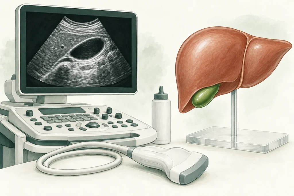
Gallbladder ultrasound helps identify stones, signs of inflammation, anatomical variations, structural changes and growths. It is a commonly used examination that gives the clinician a clearer view of the organ and helps guide further investigation and treatment. Contact the Center for Laparoscopic Surgery to check availability. A clinician can explain how the examination is performed, how to prepare and what the findings mean in the context of your symptoms.
Gallbladder anatomy
A gallbladder ultrasound describes the following features:
- location;
- size;
- shape;
- wall thickness;
- internal contents.
Interpreting the findings requires an understanding of the organ’s anatomy. The gallbladder usually lies in its fossa on the underside of the liver, between liver segments IV and V, and is attached by connective tissue. Anatomical variations matter when interpreting images and planning surgery.
The gallbladder may be oval or pear-shaped; both forms can be normal. Its main parts are the fundus, body and neck. Folds and an S-shaped configuration are anatomical variations, so the clinician looks for other findings before linking them to cholecystitis or a motility disorder. Less common variations, including duplicated ducts, can make the images more difficult to interpret and may affect diagnostic planning.
The fundus is the most distensible and mobile part; the body and neck are more closely attached to the liver. The neck continues into the cystic duct. A normal gallbladder wall is thin. The organ contracts after food, particularly fatty food, so it is usually easier to examine when fasting and filled with bile.
Gallbladder wall thickness is an important finding, but cannot establish a diagnosis by itself. In a fasting patient, a threshold of approximately 3 mm is commonly used. Measurements are generally taken at the anterior wall, which is easier to see. Bowel adjacent to the posterior wall may obscure its borders. Thickening can occur in conditions other than cholecystitis and must be interpreted alongside other findings and symptoms.
Adult gallbladder size varies with filling, the timing of meals, age and individual anatomy. If the organ is enlarged, the clinician compares the measurements with other findings rather than attributing the change to age or sex alone. Size by itself cannot confirm gallstones. When assessing their likelihood, the clinician considers that stones are more common in women and that obesity, substantial weight changes and rapid weight loss also increase risk. The size of these differences varies between the populations studied.
Approximate fasting measurements include a length of 6-10 cm, a transverse diameter of around 3 cm and a volume of roughly 30-50 mL. These reference values help describe the organ. The clinician also considers how the measurements were taken and the clinical situation, interpreting each number alongside the other examination findings.
The examination also assesses the gallbladder’s contents. Normal bile is anechoic: it reflects very little ultrasound and appears uniformly dark, usually black, on the monitor.
Preparing for an ultrasound
Before a planned scan, ask the imaging service for its preparation instructions. If bloating is a problem, a clinician may suggest adjusting your diet for a few days to improve the view. Whether changes are needed, and for how long, depends on your circumstances. Acute pain requires timely assessment: an urgent examination should go ahead without waiting to complete a preparation diet.
Depending on the examination protocol and your food tolerance, the clinician may advise you to:
- avoid alcoholic drinks;
- check whether coffee and caffeine-containing products need to be restricted;
- avoid heavy, fatty meals beforehand;
- temporarily reduce foods that cause gas for you, such as potatoes, cabbage, legumes or white bread;
- eat smaller portions if they are more comfortable to digest.
Gas in the bowel can make the gallbladder harder to see. If bloating is significant, ask a clinician whether Espumisan, Plantex or another anti-gas product is appropriate before the examination. These should not be started solely for the scan without advice. Festal, Panzinorm and Mezim are also used for specific clinical indications, rather than as a routine preparation for every patient having gallbladder ultrasound.
The day before your scan, choose familiar foods that you tolerate well, such as vegetables or porridge made with water. The timing of your last meal is calculated from your appointment and the imaging service’s instructions, rather than a fixed evening cutoff for everyone. You may be asked not to eat for at least six hours, sometimes longer. Confirm the exact interval in advance, particularly if you have diabetes or take regular medicines. A cleansing enema, including for constipation, is not normally needed for a standard gallbladder scan.
Scans are often performed in the morning after fasting. Ask the imaging service whether and when you may drink water, as protocols vary. For a meal-stimulated examination, bring the specific meal recommended by the clinician rather than an arbitrary snack.
What ultrasound shows in gallbladder disease
The gallbladder’s structure and function make it susceptible to inflammation, stones and various growths. Ultrasound helps identify these changes and determine whether further investigation is needed.
Ultrasound is generally the first imaging test for suspected gallbladder disease. Accuracy varies with the condition, examination circumstances and operator experience. Suspected acute cholecystitis or bile duct obstruction requires urgent medical assessment and sometimes additional tests.
The clinician uses an ultrasound transducer, often operating at approximately 1-5 MHz, and adjusts the settings for the patient. Images are taken in several planes and body positions, using areas that provide the clearest view, known as acoustic windows. Examining you first on your back and then on your left side helps show whether the contents move and provides a closer view of the gallbladder neck, where stones may be lodged.
Cholecystitis
Cholecystitis is inflammation of the gallbladder. It is most often related to a stone blocking the cystic duct; bacterial infection can develop, but is not necessarily the initial cause. Ultrasound helps identify stones and signs of inflammation. Obstruction, distension and impaired blood supply can contribute to ischemia. Examining the bile ducts helps investigate obstructive jaundice. Small stones sometimes lack a clear acoustic shadow, making them difficult to distinguish from sludge or a polyp.
In cholecystitis, the wall may thicken, for example to 4 mm or more. Stones, sludge, surrounding fluid and other changes may also be present. Thickening alone is insufficient for diagnosis: symptoms, physical examination and laboratory findings must also be considered. An anechoic rim, surrounding inflammatory changes or an abscess may indicate an acute or complicated process. A sonographic Murphy sign means focal tenderness when the probe presses over the gallbladder; a healthcare professional assesses this sign.
In chronic cholecystitis, the walls may become fibrotic or thickened, and the shape or size of the gallbladder may change. Folds or poorly defined outlines may also be visible. These findings are interpreted together with symptoms and other results, because enlargement is not always present and no single feature confirms the diagnosis. Inflammation involving the outer serosal surface and nearby tissues is called pericholecystitis. Marked wall thickening, including measurements of 6-10 mm, prompts investigation of its cause and assessment of the other diagnostic findings.
Polyps
Polyp-like gallbladder lesions can be found even in younger people. Most are benign, often cholesterol polyps; adenomatous lesions require a separate risk assessment. On ultrasound they usually appear as lesions attached to the wall that do not move when the patient changes position and do not cast an acoustic shadow. Nonetheless, distinguishing them from other findings on a single image is not always straightforward.
Polyps may be single or multiple. The clinician measures them and compares the results with earlier scans, considering their size, shape, growth and the patient’s risk factors. This guides follow-up: some small polyps need no surveillance, others require repeat ultrasound, and suspicious or larger lesions need a surgical opinion. Changes in echogenicity and structure are assessed alongside these features. The timing of a follow-up scan, including whether a four-month interval is appropriate, is chosen individually. Decisions about surgery also take specialist guidance into account.
With polyps, the gallbladder wall may retain its normal thickness and smooth outline. The report may therefore describe no other abnormality. The clinician still assesses the size, shape and other characteristics of the lesion itself when deciding whether further observation or treatment is needed.
Gallbladder ultrasound with functional assessment
Ultrasound can assess not only anatomy but also changes in gallbladder volume after a meal. Abnormal contraction is sometimes considered a possible contributor to symptoms, but this measurement alone cannot establish the cause of a person’s complaints.
The term “dyskinesia” refers to motility disorders of the biliary system, which can occur in younger people as well. If the symptoms and investigation findings suggest a need to assess gallbladder contraction, a clinician may request ultrasound after a meal. The examination addresses a specific diagnostic question, so it is arranged according to individual need; the test itself does not prevent other diseases. The described protocol has three stages:
- baseline measurements while fasting;
- eating an agreed meal that stimulates gallbladder contraction;
- several repeat measurements, for example three, after the meal according to the service’s protocol.
The examination may take about an hour. The clinician measures volume at specified intervals, for example every 15 minutes, and calculates the percentage change from baseline. The number of measurements depends on the protocol. Results may suggest reduced or increased contractility, but diagnosing a functional disorder also requires assessment of symptoms and exclusion of other causes, sometimes with different tests.
Contact the Center for Laparoscopic Surgery to ask about standard gallbladder ultrasound and functional assessment. At a consultation, the clinician can explain the findings, discuss treatment options and give dietary advice. Confirm current prices and appointment terms, including whether a free consultation is available, with the administrator at +380953566412. Timely diagnosis helps guide the next treatment step. Where surgery is indicated, the clinician can explain whether a minimally invasive operation is suitable.
Related reading: Gallstone types and treatment · Gallbladder inflammation · Gallbladder pain.