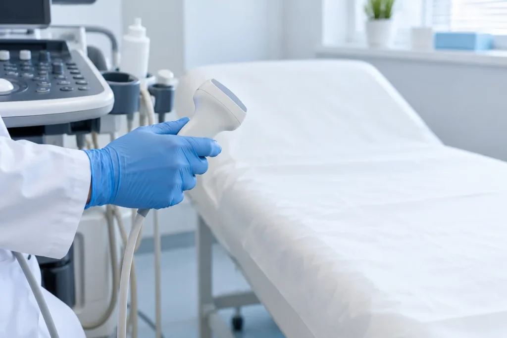
Gallbladder wall thickening is one of those phrases in an ultrasound report that can easily be mistaken for a diagnosis. In fact, it describes a finding. The cause may lie in the gallbladder itself, the conditions of the examination or the person’s overall health. Wall thickness alone is not enough to prescribe an antibiotic or recommend surgery.
First, the conditions of measurement are checked
After a meal, the gallbladder contracts and its wall may appear thicker. A planned examination is therefore usually performed after fasting according to the facility’s instructions. Gallbladder distension, the measurement site and image quality also matter. A review of ultrasound diagnosis specifically highlights the risk of apparent thickening in a contracted gallbladder. Source: review of causes of wall thickening.
If the examination was performed after eating, tell your doctor. Sometimes a properly prepared repeat ultrasound resolves the question. However, severe pain or fever must not be left untreated just to complete the required number of fasting hours. General preparation guidance is available in our article on gallbladder ultrasound.
Diffuse and localised thickening are different situations
Diffuse means that changes affect a substantial part of the wall. Focal, or localised, refers to a particular area. The doctor assesses symmetry, preservation of wall layers, contours and accompanying findings. The distinction matters when choosing further investigations, but does not establish whether a change is benign or malignant on its own.
The 2025 Korean Society of Abdominal Radiology recommendations discuss incidentally detected thickening separately from symptomatic conditions. This is an important limitation: an approach for a person without symptoms should not automatically be applied to acute pain. Source: KSAR 2025 recommendations.
An indeterminate localised change needs clarification, not a conclusion that “this is definitely inflammation” or “this is definitely a tumour”. A specialist may recommend expert ultrasound or another imaging method depending on the question that remains unanswered.
When the cause lies outside the gallbladder
The wall may become oedematous in liver disease, heart or kidney failure, and with low blood protein levels. This does not mean that everyone with this finding has one of these conditions. Rather, known diagnoses and other symptoms need to be taken into account.
A clinical study of gallbladder oedema describes cases in which systemic disease was mistaken for acute cholecystitis. The authors stress assessing the entire clinical picture to avoid unjustified intervention. Source: study of misdiagnosed oedema.
A separate prospective study in people with heart failure supports an association between wall thickness and congestion. If there is leg swelling, breathlessness or known heart disease, this information is therefore relevant to the assessment of abdominal ultrasound too. Source: study of congestion in heart failure.
How suspected cholecystitis is assessed
The doctor considers the location and duration of pain, examination findings, temperature, laboratory results and the combination of imaging signs. Stones, surrounding tissues and other changes matter as well as the wall. Read more about this symptomatic condition in “Acute cholecystitis”.
The absence of stones does not allow you to rule out a dangerous condition on your own. Equally, a stone alongside a thickened wall does not remove the need to consider other possible causes.
A practical checklist before the appointment
- Record when you last ate before the ultrasound and why it was requested.
- Bring the full report, images and earlier investigations.
- Report pain, fever, vomiting, jaundice, breathlessness and swelling.
- Prepare a list of known conditions and medicines, including recent treatment changes.
- Ask whether the thickening is localised or diffuse and how reliably it was assessed.
- Clarify what needs checking next and when to return with the results.
How to assess changes over time
A comparison should consider not only millimetres, but also the conditions of both examinations and how you felt between them. Ask the doctor whether the difference is clinically significant. Do not start treatment merely because the new report uses different wording. Keep reports in date order together with brief notes about symptoms and prescriptions.
When to seek urgent care
Severe persistent abdominal pain, fever or chills, jaundice and repeated vomiting require prompt medical assessment. Call emergency services for fainting, confusion or severe breathlessness. Do not wait for a planned repeat ultrasound. Source: NIDDK symptoms of gallstone complications.
Frequently asked questions
Does one increased measurement mean chronic cholecystitis?
No. Context, a good-quality examination and clinical assessment are needed.
Can the wall become thinner again?
This is possible, for example if the change is related to contraction or oedema. A doctor assesses the cause and changes over time.
Is a CT scan always necessary?
No. The method is chosen according to the unresolved question; a good-quality repeat ultrasound sometimes provides the necessary information.
For a routine review of your report, call +38 (095) 356-64-12. This information does not replace an individual examination.