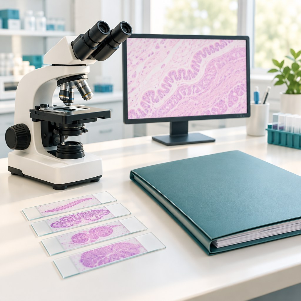
After gallbladder removal, you may receive a histology report containing unfamiliar words. “Chronic cholecystitis”, “fibrosis” or “cholesterolosis” do not automatically mean cancer. However, it is important to obtain the report and discuss it with your surgeon: sometimes examining the tissue clarifies the diagnosis and changes the next steps.
What histology examines
A pathologist examines the removed tissue, including under a microscope, and reports the changes found. This serves a different purpose from ultrasound or inspection during surgery: these show the structure of the organ at a different level.
The report usually includes the patient’s details, the type of specimen, its appearance, microscopic findings and the final diagnosis. The “Conclusion” or diagnosis section is often the easiest place to start, but it should be considered together with the comments. If testing is still ongoing, the preliminary document may be supplemented. How a pathology report is structured: NCI.
Common benign findings
- Chronic cholecystitis. Signs of longstanding inflammation in the gallbladder wall.
- Acute cholecystitis. Changes typical of acute inflammation; these may coexist with chronic changes.
- Fibrosis. An increase in connective tissue, including scar tissue.
- Cholesterolosis. Accumulation of cholesterol components in cells of the gallbladder wall; the term itself does not mean cancer.
- Calculi. The medical term for stones, if these are described in the specimen received.
These terms should not be used on their own to decide whether you need antibiotics, a special diet or another operation. For example, inflammation described in an organ that has already been removed does not prove that a postoperative infection is continuing. The report is assessed alongside your symptoms, examination and other results. Approaches to examining the gallbladder are described in the Royal College of Pathologists guideline.
What dysplasia and suspicious changes mean
Dysplasia means abnormal changes in cells. It is not the same as invasive cancer, but it needs professional explanation, taking into account the degree of change and how complete the examination is. Wording such as “suspicious”, “cannot be excluded”, or a recommendation for additional tissue sections means that the finding is still being clarified. Do not turn a preliminary possibility into a final diagnosis yourself. Definition of dysplasia: NCI.
If cancer is found unexpectedly
Gallbladder cancer is sometimes discovered after surgery performed for stones or inflammation. This finding requires a timely consultation with a specialist surgeon and an oncology team. It does not automatically mean that the disease has spread or that everyone needs the same treatment.
The decision depends on the depth of invasion, the margins of the removed tissue, other characteristics and the results of further investigations. For some very early T1a tumours, gallbladder removal may be sufficient. With deeper involvement, including T1b and above, the team assesses whether further surgery and other treatments are needed. Applying these categories to your own case without specialist advice is unsafe. Treatment approaches from the American Cancer Society.
A “negative margin” means that no tumour cells were found at the examined edge. A “positive margin” means that tumour cells were present. This is an important part of planning, but no single line replaces a full assessment. The tumour’s grade is also not its stage: these are different characteristics. About tumour grade: NCI.
How to prepare to discuss the result
- Check the surname, operation date and organ name in the report.
- Ask for a complete copy, including any addenda.
- Bring your discharge summary, operation report and previous investigations.
- Ask whether the result is final and whether the slides need to be reviewed.
- Clarify the next step, its timeframe and the doctor responsible.
Frequently asked questions
How long does histology take?
The time depends on the laboratory and whether additional tests are needed. Ask your hospital for the expected date. A delay alone does not confirm a serious diagnosis, but you should follow up on the result.
Is a second opinion needed?
It may be helpful for a complex or unexpected finding. Discuss transferring the slides and paraffin blocks to another pathologist with your doctor: a photograph of one line is usually not enough for a review.
Does benign histology explain any pain after surgery?
No. It describes the organ examined, but does not exclude postoperative complications or other causes of symptoms. New symptoms need to be assessed separately.
What if I have not received the report?
Contact the hospital where the operation was performed and ask where the specimen was sent, whether the result is ready and how to obtain a copy. Arrange for someone to explain it. Do not assume that the absence of a phone call means the report has already been checked and no further action is needed.
When not to wait for a routine consultation
After surgery, seek urgent help for increasing pain, a high temperature, jaundice, pus draining from a wound or a marked deterioration in your condition. Breathlessness or chest pain requires emergency care. Do not wait for the histology report before reporting these symptoms. Postoperative warning signs: NHS.
For a consultation about your results and next steps, call +380953566412. Read more about gallbladder removal.
This article explains general concepts and does not replace an individual assessment of your histology report.
Read also: recovery after cholecystectomy and pain after surgery.