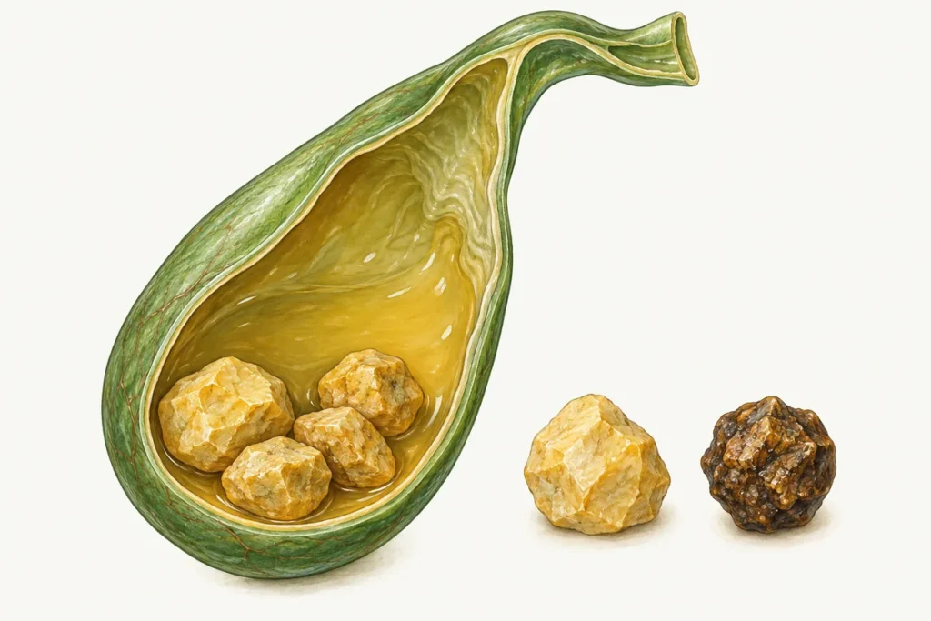
Gallstones form when the composition of bile changes or the gallbladder does not empty properly. Infections and metabolic disorders can play a role in some situations. Pain under the right ribs is a possible symptom, but many gallstones cause no symptoms at all. Treatment depends on the clinical situation: silent gallbladder stones often need no intervention, while symptoms or complications may make surgery appropriate. Persistent blockage of the bile ducts needs timely medical care.
Causes
In gallstone disease, also called cholelithiasis, substances in bile can form crystals, a sediment known as sludge, and stones of different sizes. Their development varies: small particles do not necessarily progress to large stones. Gallstones are more common in women, and pregnancy and hormonal factors can affect the risk. Their frequency also changes with age and differs between populations.
Factors and associated conditions considered during assessment:
- diabetes and other metabolic conditions; thyroid disorders are considered in the context of overall health;
- changes in bile composition and the balance of bile acids;
- an imbalance of cholesterol, bile salts and phospholipids that affects how substances remain dissolved in bile;
- excess calorie intake and an unbalanced diet; alcohol should not be used to prevent or treat gallstones;
- liver disease; pancreatic conditions may accompany bile duct problems and need their own assessment;
- some parasitic infections can affect the bile ducts, but they are not the usual cause of most gallstones;
- impaired gallbladder emptying, inflammation or infection of the bile ducts; chronic and autoimmune conditions are assessed individually;
- some types of hormone treatment;
- tumours or enlarged lymph nodes may compress the bile ducts: this is another possible cause of obstruction that needs to be distinguished from stones;
- obesity, rapid weight loss and prolonged fasting; weight changes should be gradual rather than based on extreme diets.
Excess weight, metabolic conditions and low activity levels may increase the risk of stone formation. Allergies alone do not predict whether stones will develop. To decide whether investigation is useful, the clinician considers your symptoms and overall health. Regular ultrasound is not necessary for every person who has no symptoms but is concerned about these risk factors.
Children can also develop gallstones. Possible factors include inherited susceptibility, congenital biliary abnormalities and other medical conditions. A child needs an individual assessment by a paediatrician or appropriate specialist.
Symptoms
Gallstones may remain symptom-free for a long time. They are often found incidentally on an ultrasound performed for another reason. Blood tests help assess inflammation and liver or bile duct problems, but do not replace imaging of the stones.
How you feel depends on whether a stone interferes with bile drainage and whether inflammation has developed. The clinician therefore assesses your current symptoms and investigation results, allowing for differences in how the disease affects individual people.
Possible symptoms and warning signs:
- pain, heaviness or discomfort in the upper right abdomen, sometimes after a large or fatty meal;
- biliary colic: severe upper abdominal pain that may spread to the back or right shoulder and last for hours; severe or persistent pain needs medical assessment;
- bloating, diarrhoea, belching or heartburn may occur, but are not specific to gallstones;
- fever or chills may indicate a complication;
- a bitter taste in the mouth does not establish a diagnosis;
- nausea and vomiting, especially repeated vomiting, need assessment alongside other symptoms;
- jaundice: yellowing of the skin or whites of the eyes;
- weakness and fatigue;
- dark urine and pale stools may indicate obstructed bile flow;
- chest pain, breathlessness or sudden anxiety should not be attributed to the gallbladder without assessment: urgent evaluation may be needed, including to exclude a heart problem.
An attack sometimes follows a large or fatty meal, but it may begin without an obvious trigger. The number, size and shape of the stones provide only part of the clinical picture. Tell the clinician if pain followed alcohol, stress or exertion; any connection is assessed alongside the rest of your symptoms.
For recurring symptoms, see a clinician who can decide which tests are needed and discuss treatment and diet. If pain is severe or lasts several hours, or occurs with fever, jaundice or repeated vomiting, seek urgent care rather than waiting for a routine appointment.
Diagnosis
Stones may be found during investigations for pain or by chance when there are no symptoms. Detecting them does not require waiting for severe symptoms or complications. The clinician decides which tests are useful after discussing your concerns and examining you. If you have no symptoms, the value of preventive testing is assessed individually.
Who treats gallstones? Initial assessment may be carried out by a family doctor or gastroenterologist. A surgeon assesses the need for an operation, and stones in a bile duct may require endoscopic treatment.
Assessment starts with an examination and medical history. Consider whether close relatives have gallstones, when your symptoms began, exactly where the pain occurs and how long an attack lasts. Tell the clinician about your diet, lifestyle, work, other medical conditions and medicines.
Investigations that may be used when appropriate:
- ultrasound of the liver and gallbladder is the main initial imaging test for gallstones and is usually combined with blood tests;
- an abdominal X-ray shows only some calcified stones and is not the main test for finding them;
- CT or MRI, including magnetic resonance cholangiopancreatography (MRCP), may answer further questions; endoscopic ultrasound may be used when needed;
- endoscopic retrograde cholangiopancreatography (ERCP) is mainly used when a bile duct problem needs treatment; intravenous cholangiography is not a standard first investigation;
- percutaneous transhepatic cholangiography is a specialised invasive investigation performed with imaging guidance for particular indications;
- needle-based procedures involving the intrahepatic bile ducts are carried out only on the advice of a specialist team, rather than as routine gallstone tests;
- scintigraphy may help assess biliary function or drainage in selected situations.
Complications
A complication can develop when a stone blocks a bile duct or the pancreatic duct. However, many people with symptom-free stones never develop a dangerous condition. Your clinician can help assess the risk and explain which signs of obstruction or infection mean you should seek medical help promptly.
Possible complications of gallstone disease:
- obstructive jaundice;
- acute pancreatitis and, in severe cases, pancreatic tissue necrosis;
- gangrenous cholecystitis: death of gallbladder tissue;
- cholangitis: inflammation or infection of the bile ducts;
- bile peritonitis;
- perforation of the gallbladder wall or gallbladder hydrops;
- liver damage from a prolonged blockage of the bile ducts; longstanding bile stasis can lead to secondary biliary cirrhosis, whereas a raised bilirubin result alone does not establish that diagnosis;
- a biliary fistula;
- scar-related narrowing of the bile ducts following inflammation or other injury.
Treatment
Management depends on symptoms, complications and the condition of the bile ducts. If stones are found incidentally in an otherwise normal gallbladder and cause no symptoms, observation and advice about warning signs are often sufficient. Not everyone needs medicine or surgery. Stones in the common bile duct are assessed separately.
Approaches that do not involve gallbladder surgery:
- General measures. Diet, medicines only when prescribed and follow-up tailored to symptoms and investigation results can help organise day-to-day care. They do not reliably dissolve stones or protect against every possible complication.
- Conservative care. Prescribed medicines may relieve symptoms, but ordinary pain relief does not remove gallstones. Inflammation or another complication requires its own medical management.
- Specialist options. Drug dissolution of selected cholesterol stones or lithotripsy is considered only in particular cases; neither is a universal substitute for surgery. Stones in a bile duct may be removed endoscopically.
Diet with gallstones: what to limit or adjust to your tolerance
- choose ready-made sauces, margarine and convenience foods according to their ingredients and fat content; vegetable oil can be included in an amount appropriate to your overall diet;
- very fatty meat broths may trigger discomfort, so reducing their fat content may help;
- limit large amounts of fatty meat, lard and fried food; fish and eggs are not forbidden for everyone, and portion size and preparation matter;
- sorrel, spinach, mushrooms and cabbage can be included if you tolerate them well; reduce them if they cause noticeable discomfort;
- onions, rhubarb and radishes need to be excluded only if they trigger symptoms or there is another medical reason;
- consider the fat and salt content of offal, smoked foods, canned meat and sausages;
- choose caviar, fast food and sushi with attention to ingredients, fat content, portions and food safety;
- adjust spices and seasonings to your tolerance;
- limit rich pastries, baked sweets and cream desserts as foods that may be high in fat or sugar;
- instead of frequent fried meals, consider boiling, steaming or baking with a modest amount of fat;
- chocolate and ice cream can be high in fat and sugar; fresh fruit and berries can be part of a balanced diet;
- consider your tolerance of coffee; alcohol is not a treatment and may be unsuitable with other conditions or medicines;
- use salty or high-fat cheeses in moderation and choose flavours you tolerate comfortably;
- choose dairy products according to fat content and individual tolerance;
- salt intake should follow healthy-eating advice and any requirements related to other conditions;
- the amount and type of fat should fit your nutritional needs; fat should not be eliminated completely.
If fatty meals cause discomfort, try boiling, steaming, baking or stewing with a moderate amount of fat. Choose a texture and temperature that make food comfortable to eat. Pureeing every meal or keeping stewed vegetables to a rigid weekly schedule is usually unnecessary. Let your tolerance guide these choices while keeping the diet varied and nutritionally adequate.
A balanced diet can include vegetables, fruit, wholegrains and protein sources such as lean meat and fish. Use fats in moderation, favouring unsaturated types. Butter does not prevent cholesterol deposits. Choose a regular eating pattern that suits you, and try smaller portions if large meals cause discomfort. Fluid needs depend on your circumstances, including heart or kidney conditions, so two litres is not a compulsory target for everyone. If weight loss is appropriate, aim for gradual change.
Mineral waters such as Borjomi or Essentuki are not established ways to dissolve or expel gallstones. Heating the water and drinking it before meals does not replace assessment and treatment. Discuss suitable amounts of fluid and mineral water with your clinician, taking account of sodium content and any advice for other health conditions.
For symptomatic gallbladder stones, laparoscopic cholecystectomy is often recommended. Stones in the common bile duct may require ERCP or surgical duct clearance. Laparoscopy is a minimally invasive approach, but it carries risks like any operation and is not required for every patient.
After an uncomplicated operation, discharge may be possible on the same or following day. The team assesses how you feel and the details of the procedure, then explains when to start drinking, eating, walking and resuming daily activities. Pain relief and dietary changes are tailored to your recovery; a strict diet for one or two months is not needed by everyone. Stones cannot form in the removed gallbladder, although retained or new stones in the bile ducts remain possible.
Questions and answers
Pigment stones and heavily calcified stones generally do not respond to drug dissolution. Whether an intervention is needed depends on symptoms, complications and the location of the stone, rather than simply on its hardness.
Some small cholesterol stones can shrink with specifically prescribed medicines in carefully selected patients. The decision depends on the stone and the condition of the gallbladder. Diet alone does not reliably dissolve stones, and monitoring is needed to assess how treatment is working.
Contact litholysis is a specialised method in which a dissolving substance is introduced directly into the gallbladder. It is used rarely because its risks and the characteristics of the stones must be carefully assessed. This is not a treatment to attempt yourself. A specialist team determines suitability and explains the expected result. Stones may recur after gallbladder-preserving treatment, even when dietary advice is followed.
Stone fragmentation is used only in selected situations. It is important to distinguish rarely used gallbladder lithotripsy from specialist treatment of difficult bile duct stones.
Shockwave lithotripsy uses waves generated by a machine to break a stone into fragments. It is rarely used for gallbladder stones, sometimes alongside medicines to dissolve them. The specialist considers stone composition, size and number, the condition of the gallbladder and other findings. This assessment determines whether treatment is suitable and how many procedures may be needed.
Shock waves and the resulting fragments can damage tissue. Pain, blocked ducts, inflammation and other complications are also possible. Before the procedure, the team assesses blood clotting, pregnancy, cardiovascular conditions, active inflammation and other relevant factors. This review helps identify an appropriate approach for the individual patient.
Laser lithotripsy may be used by a specialist team for selected difficult stones, particularly within bile ducts. Suitability depends on the stone and its location, and fragments can still cause complications. Access, risks and suitability are assessed individually; age or weight alone does not replace a full assessment.
Stones may recur if the gallbladder is retained. The chance of recurrence depends on the method and clinical situation.
For symptomatic gallbladder stones, the standard surgical approach is removal of the gallbladder. Removing only the stones while retaining the organ is not a routine equivalent option for every patient. This is different from removing stones from a bile duct.
Laparoscopic cholecystectomy is performed under general anaesthesia through small incisions. Carbon dioxide creates working space, and a camera guides the surgeon’s movements. The cystic duct and blood vessels are secured before the gallbladder is removed. Cholecystolithotomy has a different purpose: removing the stones while retaining the organ. The duration of either procedure depends on the approach and complexity; the surgeon can explain the expected timing for your case.
Obesity, heart disease, breathing problems, large stones or adhesions from previous operations can make treatment more complex, but they are not all absolute contraindications. The surgical team assesses the risks, anaesthetic plan and approach.
Ursodeoxycholic acid may be considered for selected small cholesterol stones, particularly when surgery is unsuitable. Not all stones dissolve, and calcification reduces the likelihood of success. Do not take bile-stimulating medicines or products that encourage gallbladder contraction without medical advice, especially if obstruction is possible.
Limitations of drug treatment:
stones can form again after treatment ends;
side effects, including diarrhoea, may occur;
treatment may take a long time;
the cost and need for monitoring should be discussed beforehand.
Dissolution may take months or years and requires medical monitoring, including ultrasound. Suitability depends on stone composition, gallbladder condition, symptoms and other illnesses. During pregnancy or with other medical conditions, a clinician must decide whether a medicine is appropriate.
Stone size alone does not determine whether an operation is needed. Symptoms, complications, the gallbladder and duct findings, and overall risk matter. Stones of different sizes can cause problems, including obstruction.
Large stones are assessed together with the gallbladder wall and your symptoms. A large size does not necessarily mean tissue necrosis, just as a small size does not rule out complications. The decision about surgery therefore takes account of the whole assessment rather than one measurement on an ultrasound report.
A small stone sometimes passes into the bowel, but this cannot safely be predicted from its size. Migration can cause obstruction, cholangitis or pancreatitis, even if pain temporarily settles. Suspected bile duct stones need medical assessment rather than waiting at home for a stone to pass.
There is no reliable, safe home method for expelling gallstones. Avoid self-diagnosis and self-treatment. Bile-stimulating products, flushes and folk remedies can make matters worse; symptoms need medical assessment.
See a clinician to assess the finding alongside your symptoms. After establishing the diagnosis, they can explain whether observation is enough or whether medicine, an endoscopic procedure or surgery is needed, and help you choose an appropriate diet. Severe persistent pain, fever or jaundice needs urgent care.
A consultation gives you time to discuss your investigation results and treatment options. The surgeon explains which procedure, equipment and anaesthetic approach are appropriate for your situation. Ask about the expected benefits, risks and alternatives so that you understand the reasons for the proposed plan.
Confirm current prices, possible concessions, accommodation and meals directly before treatment. Ask what is included in the estimate and which additional costs may arise. Contact telephone: +380953566412.
This article can help you prepare questions for a consultation and understand the available investigations and treatments. Your clinician builds an individual plan from your symptoms and test results. If signs of a complication develop, seek help promptly rather than trying to manage them with home remedies.
Related reading: Types of gallstones · Gallbladder ultrasound · Laparoscopy for calculous cholecystitis.