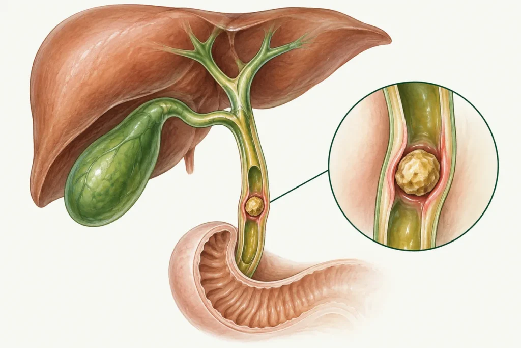
Gallstones may be found unexpectedly on an ultrasound scan or first become apparent during an episode of pain. They occur at different ages, although risk increases with age and gallstone disease is more common in women. Their formation is linked to bile composition and the way the gallbladder empties. The next step depends on symptoms. Stones that cause no problems often need no treatment. When attacks or complications occur, gallbladder removal is the main treatment, while medicines are considered for specific indications.
Why do gallstones form?
Bile contains cholesterol, bilirubin and other components. When their balance changes or the gallbladder does not empty fully, conditions can develop that favour stone formation. Stones may contain cholesterol, bile pigments and calcium salts. Understanding the cause therefore means looking at bile composition and gallbladder function, rather than simply the amount of bile present.
Several processes contribute to gallstone formation. Metabolism, associated illnesses, family history, food choices and lifestyle can all matter. During a consultation, these factors are considered together to understand the possible causes and decide on the next steps.
Possible risk factors and associated conditions are listed below. Their contributions differ, and not all are established direct causes:
- Inflammatory gallbladder or bile duct conditions;
- Metabolic disorders;
- Lipid abnormalities; high blood cholesterol is not the same as high cholesterol in bile;
- Impaired gallbladder contraction;
- Changes in bile composition and circulation;
- An unbalanced diet;
- Excessive alcohol use, particularly through its effects on the liver and pancreas;
- Smoking as a harmful general health factor;
- Certain medicines;
- Hormonal factors;
- Family history;
- Chronic cholecystitis, which may coexist with stones;
- Liver disease, including cirrhosis; the relevance of hepatitis depends on its consequences;
- Bile duct cysts or dilatation;
- Crohn’s disease, particularly involving the ileum;
- Biliary motility disorders;
- Haemolytic anaemia;
- Previous types of gastrointestinal surgery;
- Certain parasitic biliary infections;
- Cancer when it alters bile drainage or general health;
- Anatomical gallbladder or duct features affecting drainage;
- Chronic pancreatitis or the consequences of pancreatic necrosis as associated conditions needing separate assessment;
- Excess weight and obesity;
- A sedentary lifestyle.
Symptoms of gallstones
Gallstones can remain unnoticed for a long time. Symptoms are more likely when a stone obstructs bile drainage, inflammation develops or another complication occurs. The number of stones on an ultrasound report therefore does not predict the severity of pain. One small stone can cause a pronounced attack, while larger or multiple stones may cause no symptoms. The clinician considers the scan alongside how you have been feeling.
Possible symptoms requiring assessment include:
- Pain below the right ribs;
- A bitter taste;
- Loss of appetite;
- Nausea and vomiting;
- Loose stools;
- Fever when inflammation or infection develops;
- Excess intestinal gas;
- Belching;
- Heaviness below the right ribs;
- Yellowing of the skin and eyes, which needs urgent investigation.
A typical presentation of symptomatic stones is biliary colic: intense pain in the upper right or upper central abdomen. It may spread to the back, right shoulder or shoulder blade. Pain throughout the abdomen also requires assessment for other causes.
An attack may involve vomiting and pain lasting from tens of minutes to several hours. Sudden severe or prolonged pain, fever, jaundice or repeated vomiting call for urgent medical help. Do not judge the urgency only by how long a previous attack lasted: a new episode may follow a different course and needs assessment on its own merits.
Diagnosing gallstones
If gallstones are suspected, the clinician asks about pain and other symptoms, how long they last, previous health conditions and any medicines you take. After examining you, they can explain which investigations would help clarify the diagnosis. The assessment may include:
- Abdominal ultrasound;
- A full blood count and biochemical tests, including liver tests;
- Urine testing when indicated, for example to exclude another cause of pain;
- Additional biliary imaging when needed. Contrast X-ray studies are not a mandatory initial test.
Plain X-rays or cholecystography are now used infrequently. Ultrasound detects many gallbladder stones well but may miss duct stones; MR cholangiography or endoscopic ultrasound may then be appropriate.
Types of gallstones
Gallstones can be described according to several characteristics.
These include:
- Where and under what circumstances they formed;
- Chemical composition;
- Shape;
- Consistency.
The words “primary” and “secondary” describe the origin of stones in a particular context. For example, primary bile duct stones form within the ducts themselves, while secondary stones move into them from the gallbladder. If these terms appear in a report, ask where the stone is located and what that means for treatment.
By composition, stones may be described as:
- Cholesterol stones;
- Stones with substantial calcium-salt content;
- Pigment stones;
- Mixed stones.
Shape reflects composition and formation conditions. Stones may be round, oval, barrel-shaped, pointed or irregular. Some have a layered structure.
Stones range from relatively soft to hard. Treatment decisions depend more on where they are, whether they block bile drainage and whether inflammation is present. Mechanical irritation of the wall can also matter. These features are assessed together, rather than judging the risk from a single property of the stone.
Gallstone sizes
Size ranges from a few millimetres to several centimetres, with occasional stones measuring 5-6 cm. Large stones can carry certain risks, but small stones can also migrate into ducts and cause serious complications. Modern surgery can treat a range of cases, while operative risk must still be assessed individually.
Imaging may show a stone around 2 cm or much smaller stones. Size alone does not determine whether treatment is needed or which option is appropriate.
Complications of calculous cholecystitis
Stones that cause symptoms and acute inflammation can lead to life-threatening complications. Some situations require urgent surgery or endoscopic treatment. This is why new, pronounced symptoms need assessment. When stones are found incidentally without symptoms, the clinician separately considers whether observation or treatment is appropriate, rather than assuming that a complication will occur.
Possible complications or related conditions, which differ in frequency and mechanism, include:
- Acute pus-forming, including phlegmonous, gallbladder inflammation;
- Secondary liver injury and, with prolonged obstruction in selected cases, biliary cirrhosis;
- Severe impairment of liver function in appropriate complicated settings;
- Gallbladder wall perforation;
- Peritonitis, inflammation of the abdominal lining, including from bile leakage and infection;
- Gallbladder rupture;
- Intestinal obstruction, rarely caused by a gallstone;
- An abscess, a localised collection of pus;
- Cholangitis, infection or inflammation of the bile ducts;
- Sepsis;
- Blockage of a bile duct by a stone;
- Inflammatory liver changes;
- Obstructive jaundice;
- Bleeding as a rare complication;
- Acute pancreatitis;
- A fistula between the gallbladder and intestine;
- Gallbladder cancer: stones are associated with risk, but most people with stones never develop this cancer.
Complications may develop through duct blockage, infection and other related processes. Surgery performed for a clear indication reduces the risk of repeat attacks and some complications. Incidentally found stones without symptoms require a different approach: the gallbladder’s condition and any reasons for treatment are assessed first.
Treatment
Depending on symptoms and investigation results, the options may include observation, medicines or surgery. For stones that cause attacks, the standard procedure is laparoscopic cholecystectomy, which removes the gallbladder together with its stones. The clinician can explain why this option is being considered in your circumstances and what you can expect after treatment.
Medicines can relieve symptoms, and ursodeoxycholic acid can sometimes dissolve cholesterol stones. Patients are selected for this treatment using particular criteria. It takes time, does not suit every type of stone and may be followed by further stone formation. Discuss the expected benefit, limitations and alternatives before deciding whether medical treatment is appropriate.
For many patients with symptomatic stones, surgery is the better long-term option. A minimally invasive approach is often well tolerated but does not eliminate complications.
Laparoscopic cholecystectomy is performed under general anaesthesia through small incisions. The entire gallbladder is usually removed with the stones, whether they are large or small. The operation often takes around an hour, but complex cases can take longer.
Once the gallbladder is completely removed, stones cannot form within it again. However, stones can remain in or later develop within the bile ducts, so recurrent symptoms need assessment.
Potential advantages of laparoscopic cholecystectomy include:
- Smaller incisions and less tissue disruption;
- A lower wound-infection risk than open surgery, although the risk is not zero;
- Usually less blood loss than with an open operation;
- Smaller scars, rather than no cosmetic trace at all;
- Faster recovery in many uncomplicated cases.
Preparing for surgery
Preoperative preparation involves individually selected steps:
- Assessment and investigations before surgery. ECG, viral hepatitis, HIV and syphilis tests, clotting tests and a chest X-ray are included according to your health and the applicable protocol. Ask which results are needed in your case and when to provide them;
- Reviewing medicines with the clinician. Digestive enzymes for two days before surgery are not a universal requirement;
- Following the anaesthetist’s written instructions on food and fluids. A twelve-hour fast should not be independently applied as a rule for every patient.
Medicines to dissolve gallstones
Dissolution is suitable for certain cholesterol stones in carefully selected patients. The clinician considers stone composition, duct patency, gallbladder function and whether surgery is an option. Even a stone smaller than 1 cm is not enough information to choose a medicine independently; the decision requires a fuller assessment.
The following products differ in their effects and supporting evidence:
- Urolesan should not be considered a proven gallstone-dissolving medicine or used instead of indicated treatment;
- Ursosan is a brand of ursodeoxycholic acid used only for appropriate indications;
- Ursodex is an ursodeoxycholic acid product; suitability and the regimen are determined by a clinician.
If a bile acid medicine is proposed, ask about the aim of treatment and how progress will be assessed. A course can last for months or years, and the response depends on the starting situation. Stones may form again even after successful treatment. Do not begin this therapy or change the regimen without medical advice.
If attacks recur or complications develop, discuss cholecystectomy. For symptomatic stones, laparoscopic gallbladder removal is often the most effective long-term option, but the choice depends on the clinical circumstances.
Recovery advice after surgery
During recovery after laparoscopic cholecystectomy patients are generally advised to:
- Gradually return to a normal healthy diet. Temporarily limit foods that cause discomfort; a lifelong ban on all fatty, fried, smoked or spicy food is not necessary for everyone. Avoid alcohol while anaesthetic effects persist, while taking incompatible medicines and according to clinical advice;
- Start light activity and build up gradually, delaying strenuous exercise for the period recommended by the surgeon;
- Take prescribed medicines and make sure you understand the purpose of each one. Pain relief helps manage discomfort; the clinician considers the need for antispasmodics or digestive enzymes separately;
- Care for wounds and dressings according to the instructions you receive. Ask whether protective dressings are needed and when to change them. Continue to monitor healing and report any signs of possible infection, even when a dressing is in place.
Prevention
Helpful measures to reduce risk and support health include:
- A balanced diet;
- Take vitamins when there is a confirmed need. Supplements should not be used solely to prevent gallstones without a separate indication;
- Avoiding excessive alcohol;
- Maintaining a healthy weight and losing weight gradually rather than rapidly;
- Appropriate treatment of diagnosed infections and other chronic illnesses;
- Family doctor or gastroenterology reviews according to symptoms and an individual plan, rather than compulsory frequent ultrasound for everyone.
A consultation can help establish whether gallstones explain your symptoms and which investigations or treatment are needed next. Bring previous results if you have them, and note the questions you would like to discuss. Ask about costs, booking arrangements and any available free consultation on +380953566412.
Conclusion
Gallstones can remain silent, cause repeated attacks or lead to complications. The care plan depends on which of these situations applies now. Laparoscopic gallbladder removal remains the main treatment for stones that cause symptoms. Medicines have specific uses, while an incidental finding first calls for a decision about whether any treatment is needed.
Modern techniques and equipment allow minimally invasive gallbladder removal. Even a routine operation has risks, which should be discussed before consenting.
Before treatment begins, confirm the current price, which investigations and services are included, and any possible additional costs. This helps you understand the proposed plan and discuss it with the medical centre. Decisions about investigations or surgery should follow assessment of the condition, rather than depend on the price of a service alone.
Discuss the next steps based on your results and when you should seek another review. Arrange an assessment if symptoms recur. Severe persistent pain, fever or jaundice require urgent care, even if previous attacks have settled on their own.
They are hard or relatively soft formations of varying shape and size. They may be round, oval, barrel-shaped or irregular, and their composition and density differ.
Arrange a specialist consultation and bring your investigation results. If the stones cause no symptoms and the gallbladder is otherwise normal, urgent surgery is often unnecessary. Attacks or complications may lead to a recommendation for gallbladder removal or bile duct clearance. Use the appointment to understand which problem has been identified, its risks and the next step needed now.
Certain gallbladder-preserving methods exist but are not the standard equivalent alternative to cholecystectomy for symptomatic stones because of recurrence and other limitations. Duct stones are often removed endoscopically. The liver continues to make bile after gallbladder removal.
The usual procedure is laparoscopic cholecystectomy, removing the gallbladder and its stones. Ports, a camera and instruments are introduced through several small incisions in the front abdominal wall. The approach can be changed when necessary for safety.
Most people live a full life after recovery. Bile reaches the intestine without being stored first, so temporary digestive changes or diarrhoea can occur; persistent symptoms should be discussed with a clinician. Surgical complications are also possible. Most patients do not need a lifelong special diet, although individual recovery instructions matter.